Osteotomy
What Is It?
An osteotomy is a surgical procedure used to alter the alignment of the leg to change the loading pattern of the knee. A cut is made in the tibia (shin bone)and then the cut is opened or closed to change the axis of that bone.
What is it used to treat?
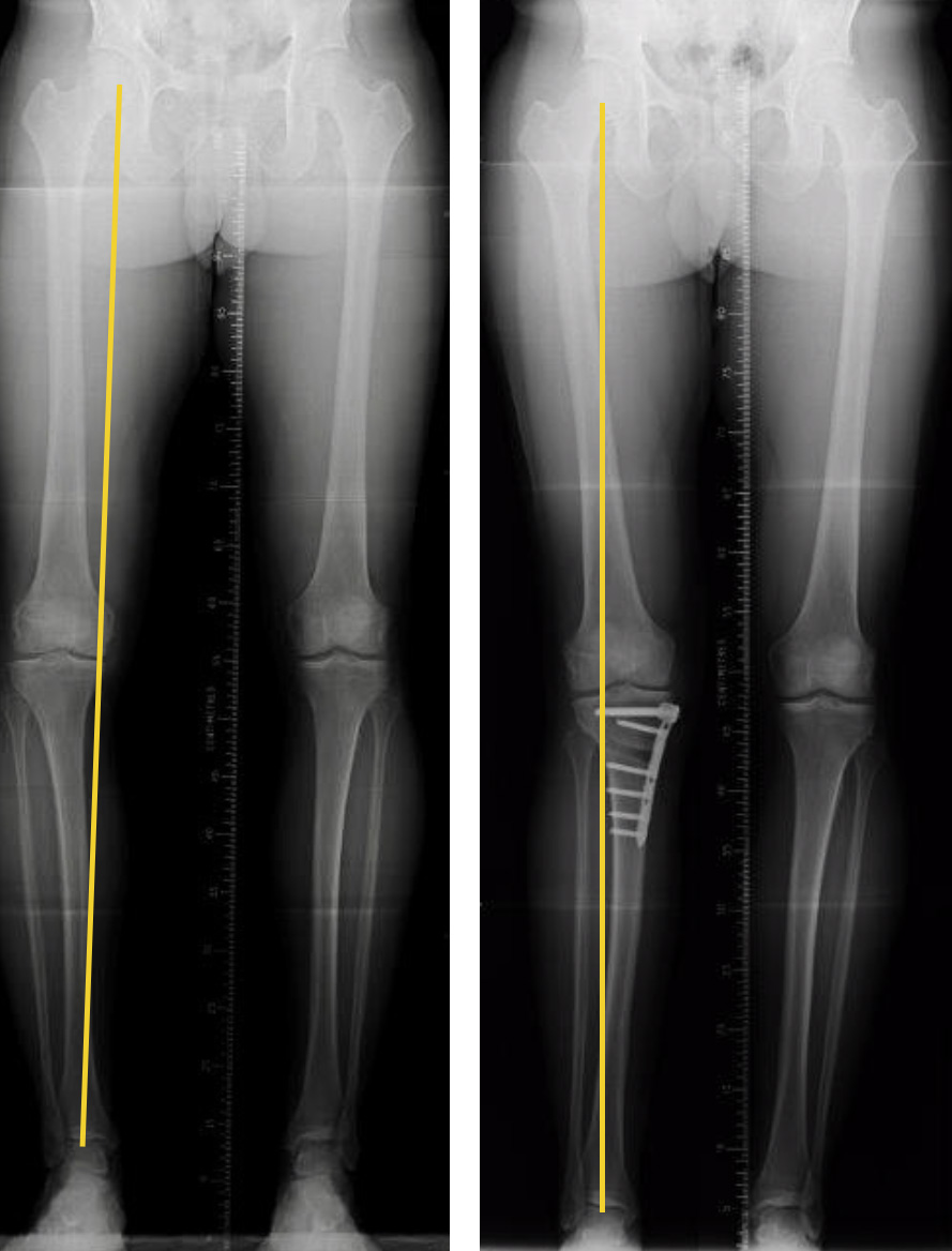
It is a procedure we use when there is wear to one side or compartment of the knee that is causing pain to that part of the knee. Most commonly it is performed when there is wear in the inner (medial) side of the knee but can be done in a slightly different way for wear in the outer (lateral) side of the knee. When the inner side of the knee is worn the leg often has a bow legged appearance (varus) because the inner side of the knee is worn down with loss of gristle (cartilage) from that side of the knee and preserved cartilage on the outer side of the knee. As a result when the affected person stands or walks more of the bodies’ weight passes through the worn part of the knee than the non worn part of the knee. That combined with the wear causes that side of the knee to be more painful and can cause further deterioration of that side of the knee. The osteotomy changes the alignment of the leg so that it is straight or very slightly aligned to the outside (knock-kneed). After the operation more of the persons weight passes through the unworn side of the knee taking some of the load off the worn side and thus decreasing the pain and slowing down the wear. The operation is a good option for patients where there is moderate wear to one side of the joint ie some but not all of the cartilage has worn away and this is causing the patient to experience regular pain. This will be demonstrated on an x-ray or MRI scan as narrowing but not complete loss of the gap between the bones (tibia and femur). When there is bone rubbing on bone in one side of the knee a partial (unicompartmental) knee replacement becomes an option and can be more successful in terms of pain relief. However in younger patients an osteotomy still remains an option
What happens in the operation?
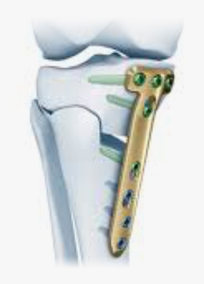
The procedure is performed under a general anaesthetic and can be performed as a day case or short overnight stay. First of all an arthroscopy (key hole surgery) is performed to assess the state of the knee joint and to ensure that it is suitable for the procedure. This means that one side of the joint is seen to be worn whilst one side of the joint is well maintained. Any damage that can be repaired via arthroscopy is attended to. The osteotomy is then performed by making a small incision on the inner side of the leg to gain access to the tibia. The inner ligament (medial collateral) is released and a controlled cut across the tibia is made using a precision saw from the inner side of the tibia towards the outer side. The bone on the outer side is left intact so that it acts as a hinge when the bone is spread apart. The procedure is performed with the surgeon using x-ray so that it can be precisely controlled. The cut in the bone is then opened up on the inside of the bone only, in effect pushing the foot end of the bone more to the outside of the leg changing the alignment in the frontal plane. A very strong plates is then fixed to the same side of the bone using screws. It bridges the gap that has been created, holding it apart at the desired correction. The plate is strong enough to take the weight of the body while the gap that has been created gradually heals up similar to a fracture in the bone.
What is the recovery like?
After the surgery some times a hinged brace is placed on the leg. These allows the knee to bend which is encouraged straight away. No plaster cast is used. The patient is usually instructed to walk with minimal weight through the leg for the first 2 weeks after the surgery. After that as much weight as the patient can tolerate can be put through the leg when walking. It can however take unto 6 to 8 weeks following the surgery before the patient can stop using crutches, very occasionally this can be up to 3 months. Most people can drive 6 weeks following the surgery. The full improvement in pain following surgery can take 6 months but most patients feel better than they did before surgery within 8 weeks.
The operation will be followed up with intermittent X-rays to check that the gap in the bone is healing up. Occasionally the plate irritates the soft tissues that lie over it and it has to be removed this is not usually done, if required, until a year after the original operation to ensure the bone is fully healed. This is a much smaller procedure than the osteotomy itself.

X-rays before and after osteotomy to demonstrate change in alignment of the leg. Yellow line demonstrates the position of load across the knee.

